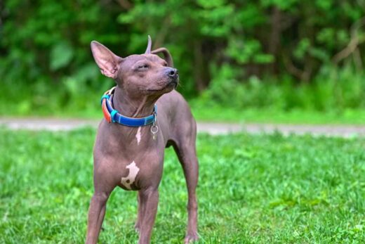
While you and I may experience an occasional cough, sneeze, or even asthma in response to environmental allergens, our pups are more likely to experience an uncomfortable skin condition called atopic dermatitis. Dermatological issues are a primary worry of many pet parents, and understanding Atopic Dermatitis can be a step in helping your pup. We want our dogs to be healthy and comfortable, so here is what you can do to help them feel their best if allergies strike.
Environmental allergens, such as dust, pollen, and mold, can cause an atopic allergic reaction or Atopic Dermatitis (AD). Atopy is the tendency to produce an exaggerated immunoglobulin E immune response to otherwise harmless substances in the environment. Immunoglobulin E (IgE) is a type of antibody found in mammals synthesized by plasma cells, and it is an important part of the immune response.
So, Canine Atopic Dermatitis (CAD) is a common skin disorder wherein a dog has a hereditary predisposition to develop an itchy inflammatory skin disease associated with elevated levels of IgE antibodies, which typically target environmental allergens. CAD is characterized by chronic itchiness and distribution of skin lesions, which are areas of skin that look different from the surrounding area (ex. bumps or patches).
CAD researched placed initial emphasis on understanding the IgE response to allergens. Elevated IgE levels are undoubtedly significant in the pathogenesis of most cases of CAD. However, research efforts have recently uncovered that the disease is multifactorial and dependent on a range of other biological factors like T-cell polarization, altered mast cell releasability, and impaired skin barrier function. There are a higher proportion of mast cells in a dog’s skin, which release histamines and other substances in the face of an allergic challenge. In humans with AD, an impaired skin barrier is an important precursor to developing the disease. It is also known that the combination of the impaired skin barrier with allergen exposure explains the development of an IgE response to the offending allergens.
Multiple gene expressions involved in skin barrier function and inflammation have been described as down- or upregulated in the skin of atopic dogs. 361 genes relevant for inflammation, wound healing, or immune response processes showed an increased expression, whereas 226 genes associated with differentiation and skin barrier function showed decreased mRNA concentrations in allergen-treated skin of sensitized dogs. This means genetic evidence suggests that dogs with AD have increased inflammation and immune response while they have reduced skin barrier function.
A dog with atopic dermatitis will usually show signs and symptoms between 3 months to 6 years of age. The condition often begins mildly, with symptoms not becoming clinically visible before the third year.